The SNE-Alpha is a desktop and tabletop scanning electron microscope by SEC Co. Ltd., distributed in the US exclusively by NanoImages. It delivers 5nm resolution at 30kV, 250,000x magnification, and reaches full vacuum in 90 seconds. 40% smaller than previous-gen tabletop SEMs — no dedicated room, no 3-phase power, no specialist operator required. Available to buy or lease from $1,500/month.
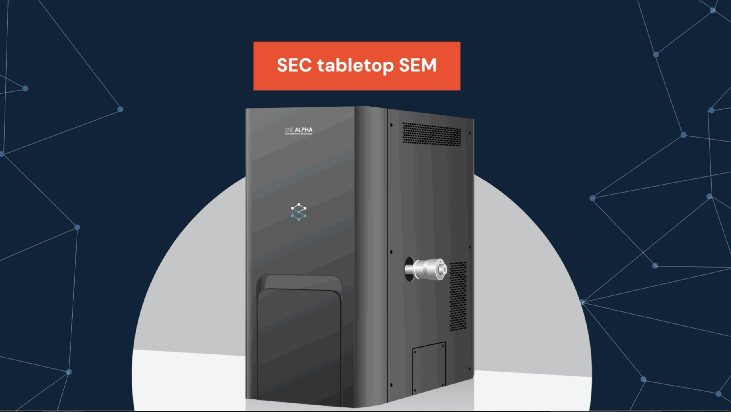
SNE-Alpha Desktop SEM
Don’t let the size fool you. Professional-grade specifications for researchers who demand clarity.
The SNE-Alpha represents the next generation of desktop scanning electron microscopes from SEC. With a 40% reduction in footprint compared to previous models—without sacrificing the column physics required for high-resolution imaging—it delivers floor-model performance in a compact benchtop package that fits any lab environment.
The SNE-Alpha pushes beyond the limits of optical microscopy with up to 250,000x magnification and 5nm resolution – allowing ultra-detailed visualization of nanostructures that would be impossible to see with traditional microscopes. Its dramatically enhanced interface displays results 60% faster than previous software, with upgraded automatic functions including newly added Auto-Gun-Align and enhanced Auto Focus for easy image capture.
Key Features
5nm Resolution
Best-in-class resolution at 30kV rivals floor-model SEMs. See nanoscale structures with exceptional clarity and detail.
90-Second Vacuum
Start imaging in under 2 minutes—50% faster than competitors. Vent time of only 15 seconds enables rapid sample changes.
5-Axis Motorized Stage
Standard motorized stage with X, Y, Z, rotation, and tilt. High-precision navigation for accurate sample positioning.
Dual Detectors Standard
Both SE (Secondary Electron) and BSE (Backscattered Electron) detectors included. Switch instantly between imaging modes.
Image Stitching
Capture a wider range with large area scans. Built-in stitching creates panoramic images of your entire sample.
3D Surface Analysis
Built-in 3D rendering functions allow inspection and analysis of surface roughness directly in the software.
Auto Functions
Auto-Gun-Align, Auto Focus, Auto Brightness, and Auto Contrast. Get optimal images with minimal manual adjustment.
Analytical Options
Factory-ready ports for EDS, Raman, EBSD, CL, and EBIC integration. Expand capabilities as your needs grow.
Technical Specifications
| Parameter | Specification |
|---|---|
| Resolution (SE) | 5nm @ 30kV |
| Resolution (BSE) | 10nm @ 30kV |
| Magnification Range | 20X to 250,000X |
| Accelerating Voltage | 1kV to 30kV (1kV steps) |
| Electron Source | Pre-centered tungsten hairpin filament |
| Detectors (Standard) | SE (Secondary Electron) + BSE (Backscattered Electron) |
| Vacuum System | Turbomolecular pump + rotary pump |
| Pump Down Time | 90 seconds to operating vacuum |
| Vent Time | 15 seconds |
| Stage Type | 5-axis motorized (X, Y, Z, Rotation, Tilt) |
| Stage Travel (X/Y) | 40 mm X 40 mm |
| Stage Travel (Z) | 40mm |
| Tilt Range | -45 to +90 degrees |
| Rotation | 360 degrees |
| Working Distance | 5mm to 40mm |
| Image Capture | Up to 5120 X 3840 pixels |
| Dimensions (W x D x H) | 300mm x 465mm x 600mm |
| Weight | ~85 kg (main unit) |
| Power Requirements | 110-240V AC, 50/60Hz, 1.5kVA |
Why Choose the SNE-Alpha?
| Feature | SNE-Alpha | Typical Competitors |
|---|---|---|
| Resolution | 5nm | 8-15nm |
| Magnification | 250,000X | 100,000-150,000X |
| Voltage Range | 1-30kV | 5-20kV typical |
| Stage | 5-axis motorized | 3-4 axis, often manual |
| Vacuum Time | 90 seconds | 2-5 minutes |
| Filament | Tungsten (low cost) | CeB6 or LaB6 (expensive) |
Optional Accessories & Upgrades
Bruker XFlash 630 EDS
Add elemental analysis with the Bruker XFlash silicon drift detector. Detect elements from Boron (B) to Uranium (U).
Raman Spectroscopy
Correlative Light and Electron Microscopy (CLEM) for simultaneous chemical and structural analysis.
EBSD Analysis
Electron Backscatter Diffraction for crystallographic orientation and phase identification.
MCM-100 Sputter Coater
Compact sputter coater for non-conductive sample preparation. Built-in rotary pump.
Resources
See the SNE-Alpha in Action
Send us your samples and we’ll show you exactly what this system can do.
Or call us: (925) 297-5668