Posts by admin
Full Size SEM Perpetually Overwhelmed: Unleash Relief And Potential With A Desktop SEM!
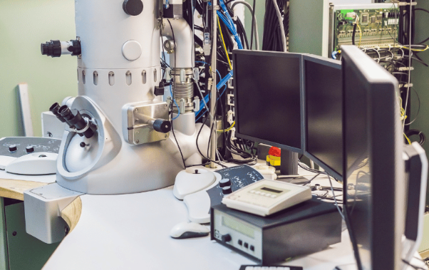
Maximizing Analytical Capabilities: The Strategic Role of Desktop SEMs in Modern Laboratories In today’s fast-paced research environment, analytical laboratories face high demands for both sample throughput and detailed material analysis. While high-end field emission SEMs remain the gold standard for advanced research, they often become bottlenecks due to heavy use and high demand. This is…
Read MoreRevolutionary Metals Research: Laser Heated EBSD Analysis with Tabletop SEM
Laser Heated EBSD Laser Heated EBSD Experiments The integration of laser heating capabilities with Electron Backscatter Diffraction (EBSD) analysis represents a groundbreaking advancement in materials science research. When combined with the SNE-Alpha Tabletop Scanning Electron Microscope (SEM) equipped with Bruker EDXS capabilities, this innovative approach offers researchers unprecedented insights into material behavior under dynamic temperature…
Read MorePolymer Research
Polymer Research:How Integrated Raman Spectroscopy Revolutionizes Tabletop SEM Analysis Understanding the Power of Combined Analytical Techniques In the dynamic world of polymer research and development, the ability to comprehensively characterize materials at multiple levels is crucial. The integration of Raman spectroscopy with the SNE-Alpha Tabletop SEM and EDS capabilities creates a powerful analytical platform that…
Read MoreTabletop SEM with Raman Spectroscopy For Graphene Research
SNE-Alpha Tabletop SEM with EDS and Raman Spectroscopy: Revolutionary Graphene Analysis Introduction: A Breakthrough in Materials Science Moreover, the characterization of graphene and its derivatives has always been a critical challenge in materials science. Now, enter the SNE-Alpha Tabletop Scanning Electron Microscope (SEM), integrated with Raman spectroscopy and Energy Dispersive X-ray Spectroscopy (EDS) capabilities. Consequently,…
Read MoreAdvancing Pharmaceutical R&D with a Tabletop SEM with Raman
Making Pharmaceutical Research Easier: A New Approach to Sample Analysis with a Tabletop SEM and Raman The Challenges Researchers Face In pharmaceutical research, scientists work hard to understand complex materials. However, their current methods make this work much harder than it needs to be. A tabletop SEM with Raman and EDS may be the answer…
Read MoreDominate Metals Research: Unlock Advanced SEM/EBSD/EDS Analysis
Understanding the Bruker QUANTAX ED-XS EBSD and EDS detector and SNE-Alpha Tabletop SEM: A Simple Guide What Makes This SEM Special? A New Way to Look at Materials In today’s world of science, researchers need tools that are both powerful and easy to use. Moreover, the combination of Bruker’s QUANTAX ED-XS with the SNE-Alpha Tabletop…
Read MoreCell Membrane Picture: Peering Inside a Living Cell Using Microscopes

Ask a cell biologist about their passion, and they might reveal a secret love for microscopes. After all, who wouldn’t be captivated by the beauty of a cell membrane picture, a window into a world teeming with life? This hidden wonder surrounds us, from the delicate patterns of a rose petal to the carrot in…
Read MoreScanning Electron Microscope for Sale: 10 Points to Consider When Making Buying Decisions
When examining a scanning electron microscope for sale, the decision-making process involves more than just selecting the most advanced equipment available. Such powerful tools, essential for detailed analysis in fields ranging from materials science to biology, demand careful consideration to contribute to the success of your work. In this blog, we’ll cover ten key points…
Read MoreSEM vs. TEM: Understanding the Key Differences and Applications
Electron microscopy has revolutionized our ability to see the world at the nano and cellular levels. Two primary techniques, Scanning Electron Microscopy (SEM) and Transmission Electron Microscopy (TEM), offer powerful ways to explore materials, biological specimens, and more. In this blog, we’ll look into the key differences and applications of SEM vs. TEM, guiding you…
Read MoreRaman Spectroscopy FAQ
A Look At Raman Spectroscopy What is it? It is a technique that analyzes the vibrational modes of molecules using light. When a laser beam interacts with a sample, most of the light scatters elastically (Rayleigh scattering). However, a small portion of the scattered light changes energy (inelastically scatters) due to interactions with the molecule’s…
Read More