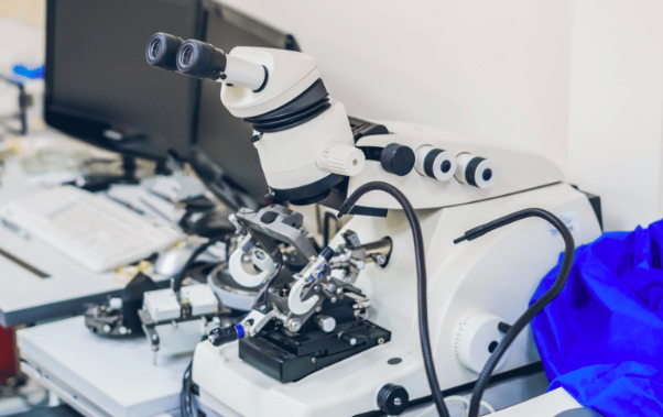
SEM vs. TEM: Understanding the Key Differences and Applications

Electron microscopy has revolutionized our ability to see the world at the nano and cellular levels. Two primary techniques, Scanning Electron Microscopy (SEM) and Transmission Electron Microscopy (TEM), offer powerful ways to explore materials, biological specimens, and more. In this blog, we’ll look into the key differences and applications of SEM vs. TEM, guiding you through their operations, unique features, and how to choose the right one for your needs.
What is SEM?
SEMs provide a way to get high-resolution images of a sample's surface. By scanning a focused beam of electrons across the specimen, these tools generate detailed images that reveal the texture, morphology, and chemical composition of the surface.
How does an SEM Work?
The process begins with an electron gun producing a beam of electrons. This beam, finely focused by electromagnetic lenses, scans over the sample in a patterned manner. As the electrons interact with the sample's surface, they generate signals like secondary and backscattered electrons. Detectors collect these signals to form an image that depicts the surface's topography and composition with remarkable clarity.
What is TEM?
TEMs allow researchers to visualize the internal structure of a specimen at the atomic or molecular level. It passes a beam of electrons through a very thin sample and captures the transmitted electrons to create an image.
How Does a TEM Work?
In a TEM, the electron beam emitted by an electron gun is focused onto the specimen by condenser lenses. The beam that passes through the sample interacts with the specimen's atoms, losing energy and changing direction. The transmitted electrons are then magnified by objective lenses and projected onto a detector, resulting in an image that reveals the internal structure of the sample.
SEM vs. TEM: Operational Differences
When choosing between SEM and TEM, understanding their operational differences is necessary. The following distinctions impact not only the type of data you can gather but also how you prepare your samples and what you can ultimately learn about them:
Imaging Techniques
The core difference between SEM and TEM lies in their imaging approaches. SEM offers the best quality images of a sample's exterior, capturing its texture and composition with depth. It does this by directing a focused beam of electrons across the surface, collecting the emitted signals to form an image.
In contrast, TEM provides a window into the sample's interior, achieving higher-resolution images that can display atomic structures. This is accomplished by sending electrons through the specimen and capturing those that emerge on the other side.
Electron Interaction
The interaction of electrons with the sample is another distinguishing factor. In SEM, the primary focus is on the electrons that are scattered off or emitted from the surface of the specimen. These interactions help form images that reveal the topography and elemental composition of the sample.
Meanwhile, TEM relies on the electrons that pass through the specimen, using the variations in this transmission to construct detailed images of the internal structure, including defects and the arrangement of atoms.
Application Suitability
The choice between SEM and TEM also hinges on the specific application at hand. SEM is the go-to technique for analyzing surface features, providing insights into the sample's external characteristics. It's beneficial for studying the morphology and chemical composition of various materials.
Conversely, TEM is indispensable for exploring the inner makeup of a specimen, from the crystallography of materials to the detailed organization within biological cells. Its ability to illuminate internal features at the nanoscale makes it a powerful tool for advanced scientific research.
Knowing how to compare and contrast a tem and a sem is crucial if you’re looking to employ electron microscopy techniques and ensure the selection of the proper method for your specific research and investigational needs.
Decision Factors When Buying an Electron Microscope
The following factors will guide you in selecting the electron microscope that aligns best with your research objectives, financial parameters, and operational requirements:
Resolution
One of the primary considerations when choosing between SEM and TEM is the level of detail you need. TEM has the upper hand for tasks requiring atomic or molecular resolution, making it suitable for in-depth analysis of internal structures. While offering excellent surface detail, SEM cannot match TEM's fine resolution capabilities for internal examinations.
Sample Preparation
The nature of your samples and the preparation they require can also guide your decision. TEM demands thinner samples to allow electrons to pass through, which might necessitate elaborate preparation methods. In contrast, SEM accommodates a broader range of sample types and thicknesses, providing a more straightforward preparation process for many applications.
Analysis Goals
Your specific analysis goals also influence your choice. If your interest lies in exploring the surface characteristics and topology of materials, SEM will likely meet your needs with its detailed surface imaging.
Conversely, if your research or quality control processes require a look at the internal structure of materials, TEM's superior internal visualization capabilities will be more beneficial.
Budget
Finally, budget constraints play a significant role. Generally, TEM systems are more costly than SEM in terms of both purchase price and operational expenses, including preventive maintenance and sample preparation costs. When budget is a limiting factor, SEM offers a cost-effective solution without sacrificing significant imaging capabilities for surface analysis.
Combining SEM and TEM Technology
The world of electron microscopy offers a powerful choice: compare and contrast a TEM and a SEM to select the right tool for your analysis strategically. However, cutting-edge instruments now seamlessly combine the strengths of an SEM and a TEM. This hybrid approach provides unprecedented flexibility, allowing researchers to switch effortlessly between SEM's detailed surface imaging and TEM's high-resolution exploration of internal structures – all within a single system.
These integrated platforms streamline research in fields like materials science, biology, and nanotechnology. No longer does complex analysis require separate instruments, making investigations more efficient and revealing a more complete picture of the analyzed samples. This technological fusion minimizes compromises and lets scientists and researchers who previously had to choose between the contrasting approaches of SEM and TEM take full advantage of their capabilities.
Key Takeaways: Choosing Between SEM vs. TEM
In conclusion, understanding the difference between SEM and TEM is crucial if you’re involved in any discipline that requires microscopic analysis.
When choosing between these powerful techniques, consider your specific needs: the resolution required, sample preparation capabilities, the focus of your analysis, and budget constraints. Advances in technology have also introduced hybrid systems, combining the strengths of both SEM and TEM, offering a versatile solution for comprehensive analysis.
Are you ready to dive deeper into the microscopic world with the right electron microscopy technique? Explore our range of electron microscopy solutions to find the perfect match for your analytical needs and unlock the full potential of your research today.
