
Biology & Life Sciences
Explore biological structures from cells to organisms with nanometer resolution SEM imaging
Scanning electron microscopy reveals the intricate surface details of biological specimens that optical microscopes cannot resolve. From the ultrastructure of cell membranes to the complex morphology of insects, SEM provides unparalleled depth of field and three-dimensional visualization of biological samples.
The SNE-Alpha desktop SEM enables biological imaging without the complexity of traditional floor-model systems. Variable pressure mode allows imaging of hydrated and delicate specimens with minimal preparation, while the compact design fits easily in teaching labs and research facilities.
Key Biology Applications
- Microbiology: Image bacteria, fungi, and biofilms at high resolution. Study cell morphology, attachment mechanisms, and antimicrobial effects
- Cell Biology: Visualize cell surface features, membrane structures, and cell-cell interactions in fixed preparations
- Entomology: Study insect morphology, compound eyes, wing scales, sensory structures, and surface textures
- Botany: Examine plant surfaces, stomata, trichomes, pollen grains, and seed coat structures
- Parasitology: Characterize parasite morphology, attachment structures, and surface antigens
- Marine Biology: Image diatoms, foraminifera, and other microorganisms with shell structures
Sample Preparation for Biological Specimens
Biological samples typically require preparation to withstand the vacuum environment and electron beam. Common preparation methods include:
- Chemical Fixation: Glutaraldehyde and osmium tetroxide preserve cellular ultrastructure
- Critical Point Drying: Prevents surface tension artifacts during drying
- Sputter Coating: Apply thin gold or platinum layer to prevent charging on non-conductive samples
- Variable Pressure Mode: Image hydrated samples with reduced preparation requirements
Biology SEM Images
Sample images captured with the SNE-Alpha desktop SEM. Click to enlarge.
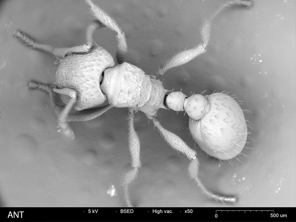
Ant
Detailed surface morphology of ant exoskeleton and appendages
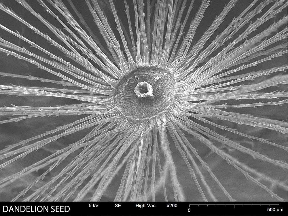
Dandelion Seed
Pappus structure showing delicate filaments for wind dispersal
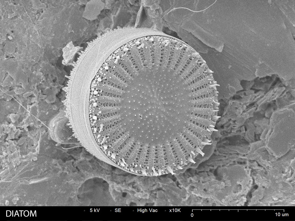
Diatom
Marine microorganism with intricate silica frustule structure
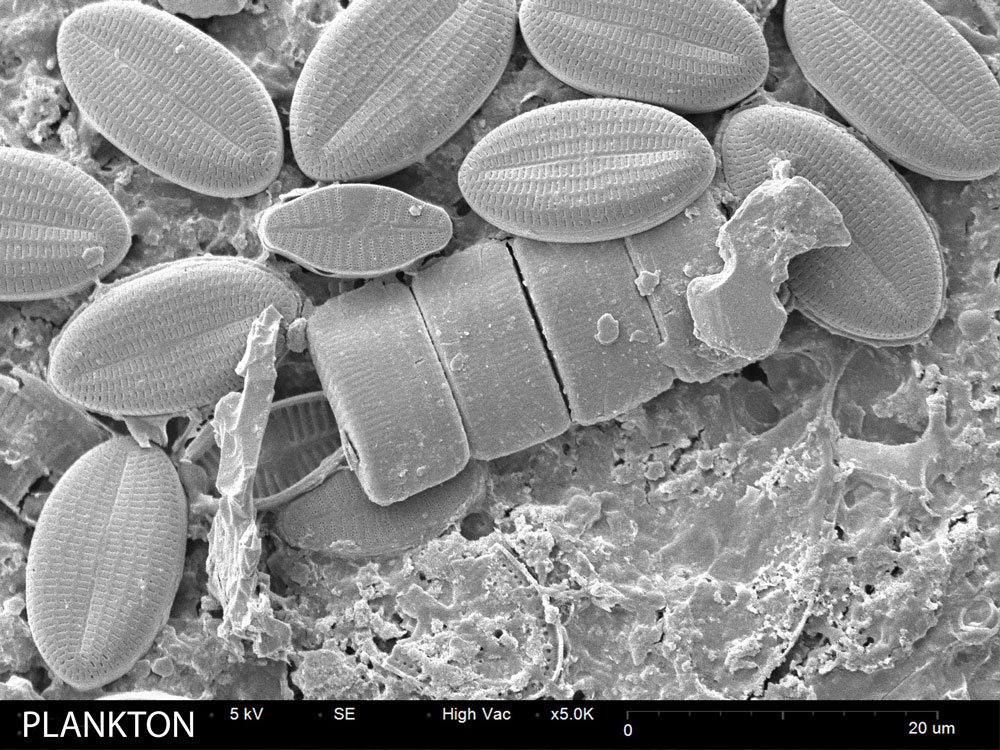
Plankton
Marine plankton showing detailed surface features
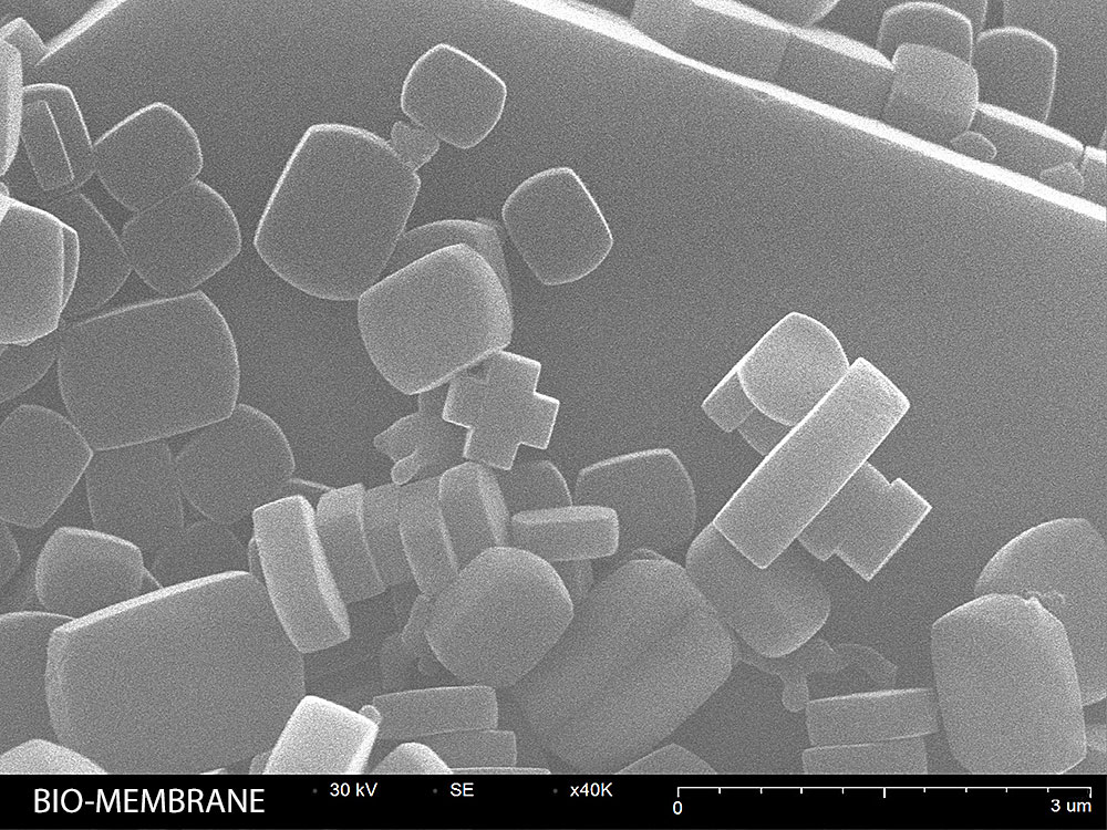
Biological Membrane
Membrane structure showing surface topology and pore features
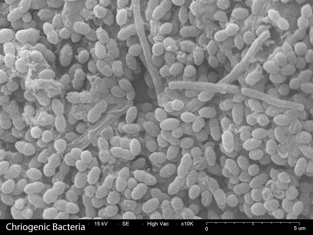
Bacterium
Bacterial cell morphology preserved via cryogenic preparation
Common Research Areas
Teaching & Education
Desktop SEM brings electron microscopy into undergraduate labs. Students can prepare and image their own samples during class.
Biomedical Research
Study biomaterials, implant surfaces, drug delivery particles, and tissue scaffolds at high resolution.
Food Science
Analyze food microstructure, starch granules, protein networks, and microbial contamination.
Veterinary Medicine
Examine parasites, pathogens, and tissue samples for diagnostic and research purposes.
Recommended Equipment
SNE-Alpha Desktop SEM
Compact, high-resolution scanning electron microscope ideal for research and quality control applications.
Bruker XFlash EDS
Energy dispersive X-ray spectroscopy for elemental analysis and material identification.
MCM-100 Sputter Coater
Prepare non-conductive samples with gold or platinum coatings for optimal SEM imaging.
Related Applications
Resources
Explore the Living World
See how the SNE-Alpha can advance your biological research and teaching programs.
Request Sample Analysis