SNE-Alpha Desktop SEM: Why Buy a Room When You Need a Microscope?
5nm resolution. 90-second vacuum. Built by microscopists, not salespeople.
Used by researchers at the following institutions
Browse 30+ published research papers using SEC desktop SEMs →
Google · Meta · MIT · UC Berkeley · Purdue University · Texas A&M · Sandia National Laboratories · Oak Ridge National Laboratory · Canadian Nuclear Laboratories
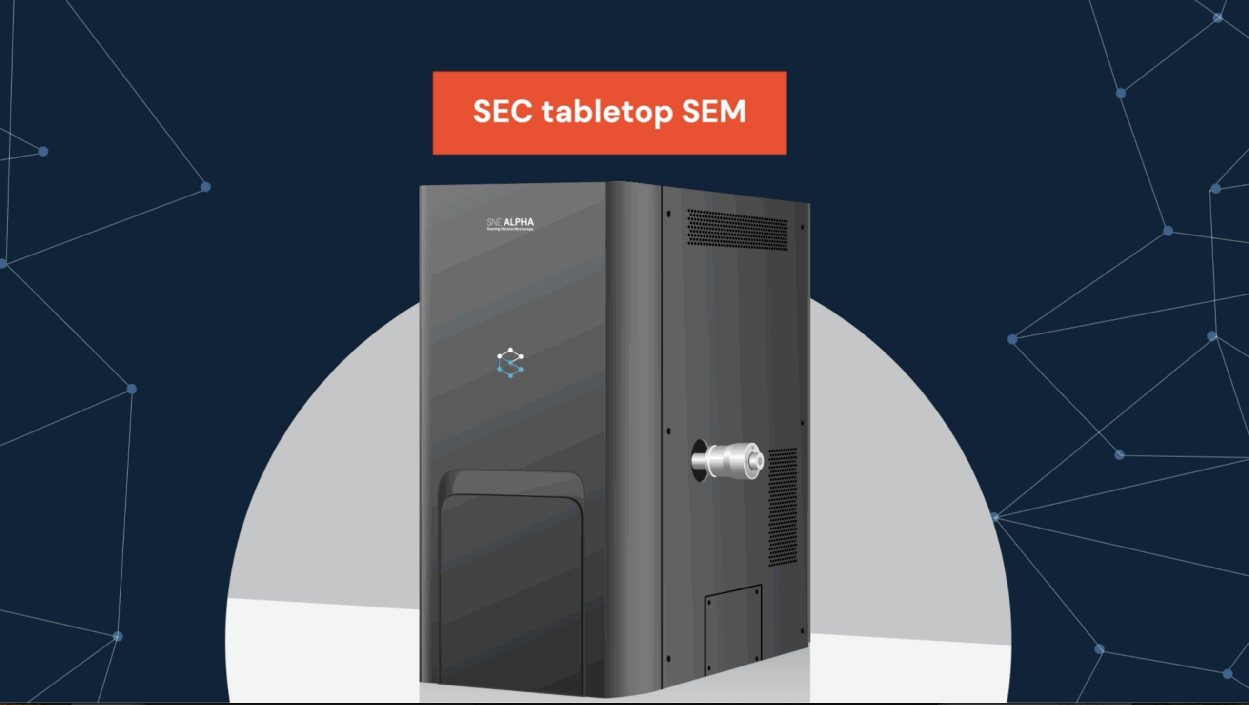
What is the SNE-Alpha Desktop SEM?
The SNE-Alpha is a benchtop scanning electron microscope that delivers 5nm resolution at 30kV and 90-second pump-down—research-grade electron optics in a footprint 40% smaller than traditional floor-model SEMs.
The SNE-Alpha is a desktop scanning electron microscope (SEM) manufactured by SEC Co. Ltd. and distributed exclusively in the United States by NanoImages. Its precision electron optics and beam deceleration technology deliver 5nm resolution at 30kV with minimal astigmatism correction needed, making it ideal for labs that lack space for traditional floor-model SEMs. The SNE-Alpha features a 5-axis motorized stage, dual SE/BSE detectors, and reaches operating vacuum in just 90 seconds—50% faster than competitors.
Key Takeaways: SNE-Alpha Desktop SEM
- 5nm resolution at 30kV — research-grade imaging in a benchtop form factor, no dedicated room required.
- 90-second vacuum, 15-second vent — fastest pump-down of any desktop SEM, enabling high-throughput sample workflows.
- Full analytical suite — integrates EDS, EBSD, Raman, and cathodoluminescence for elemental, crystallographic, and chemical analysis.
Why researchers choose the SNE-Alpha
The SNE-Alpha combines 5nm resolution, 90-second vacuum pump-down, and a full 1–30kV accelerating voltage range—delivering floor-model SEM performance on any lab bench.
Best in class for desktop SEM
Microscopist’s note: This resolution is particularly effective for imaging carbon nanotubes and nanopowders without the charging artifacts common in older desktop SEMs.
50% faster than competitors
Microscopist’s note: We routinely swap 10+ samples in an hour during pharmaceutical particle studies—the 15-second vent makes batch workflows practical on a desktop SEM.
Fits on any lab bench
From macro to nano scale
X, Y, Z, rotation & tilt
Microscopist’s note: The tilt capability is a game-changer for fracture surface analysis—we can angle the sample to reveal grain boundaries that flat-mount imaging misses entirely.
Full range for any sample
Microscopist’s note: Low kV is essential for beam-sensitive biologicals and polymers. We regularly drop to 5kV for tissue samples that would melt under standard 20kV conditions.
SNE-Alpha Desktop SEM
Floor-model electron optics in a benchtop form factor. The SNE-Alpha delivers research-grade secondary and backscatter electron imaging without requiring a dedicated microscopy suite, vibration isolation table, or special facility infrastructure.
- 1-30kV accelerating voltage (1kV steps)
- 5-axis motorized stage with <5μm accuracy
- SE and BSE detectors included
- High vacuum standard, low vacuum optional
- Optional: EDS, Raman, EBSD, CL integration
Built for your research
From semiconductor wafer inspection to forensic trace evidence, the SNE-Alpha serves 17 application areas across materials science, life sciences, and industrial quality control.
Materials Science
Metals, polymers, composites, failure analysis
Life Sciences
Cells, tissues, biological structures
Forensics
Evidence analysis, trace materials, GSR
Geology
Minerals, rocks, paleontology
Nanomaterials
Nanoparticles, thin films, quantum dots
Pharmaceuticals
Drug formulation, particle characterization
Semiconductors
Wafer inspection, defect analysis, IC packaging
Energy
Batteries, solar cells, fuel cells, catalysts
See the SNE-Alpha in action
Watch the SNE-Alpha desktop SEM cycle from sample loading to high-resolution imaging in under two minutes.

Get a Quote in 24 Hours
NanoImages provides same-day pricing for the SNE-Alpha desktop SEM, with lease options starting at $1,500/month.
Tell us about your application and we’ll send you a tailored quote with pricing, lease options, and sample imaging results.
- No obligation, no pressure
- Free sample imaging available
- Lease options from $1,500/mo
Frequently Asked Questions
Common questions about the SNE-Alpha desktop SEM—resolution, vacuum speed, detectors, EDS integration, and how it compares to Phenom.
What is the resolution of the SNE-Alpha desktop SEM?
The SNE-Alpha achieves 5nm resolution at 30kV—best-in-class for any desktop SEM. This allows researchers to image nanoscale structures with clarity comparable to larger floor-model SEMs, without dedicating an entire room to the instrument.
How long does vacuum pump-down take?
Just 90 seconds to operating vacuum—50% faster than comparable desktop SEMs. Vent time is only 15 seconds, enabling rapid sample changes and high-throughput workflows where multiple samples need imaging in a single session.
Does the SNE-Alpha require sample coating?
No—the optional low vacuum mode allows imaging of non-conductive samples without any coating. For optimal resolution on non-conductive samples, gold, platinum, or carbon coating is recommended using our MCM-100 sputter coater.
Can I add EDS for elemental analysis?
Yes—the SNE-Alpha integrates with the Bruker XFlash 630 EDS detector, covering Boron (B) to Uranium (U). Additional analytical options include Raman spectroscopy for chemical identification, EBSD for crystallographic mapping, and cathodoluminescence for defect analysis.
How does the SNE-Alpha compare to Phenom?
The SNE-Alpha offers a larger chamber, broader voltage range (1–30kV), and lower operating costs than Phenom desktop SEMs. It uses affordable tungsten filaments instead of proprietary cartridges, provides more flexible sample mounting options, and delivers comparable resolution at a lower total cost of ownership.
Latest Insights from the Lab
Pharmaceutical Analysis with SEM-EDS-Raman
How multimodal desktop SEM workflows accelerate drug formulation studies.
Raman Spectroscopy FAQ
Everything researchers need to know about integrating Raman with desktop SEM.
SEM-Raman for Radioactive Samples
Desktop SEM advantages for imaging and analyzing radioactive materials safely.
Partners & Resources
Our Partners
- SEC Co. Ltd. — SEM Manufacturer
- Bruker — EDS & Analytical Systems
- Waviks — Imaging Solutions
Learn More
Quick Links
Proof is in the Pixel
Send us your samples and we’ll show you exactly what the SNE-Alpha can do.
Schedule Your DemoOr call us: (925) 297-5668