Gunshot Residue Evidence: A Two-Part Puzzle
When a firearm is discharged, it releases a complex cloud of microscopic debris known as gunshot residue (GSR). This residue comprises both inorganic particles from the primer and organic compounds from the propellant. Traditional GSR detection relies heavily on scanning electron microscopy coupled with energy dispersive X-ray spectroscopy (SEM/EDS), which effectively identifies characteristic metal particles such as lead (Pb), barium (Ba), and antimony (Sb). However, this technique misses the organic fraction of the residue—an increasingly important component, especially with the rise of lead-free ammunition.
To bridge this gap, forensic scientists are turning to Raman spectroscopy. By integrating Raman with SEM, investigators can simultaneously detect both the metallic and organic components of GSR on a single platform, yielding a more complete and convincing forensic profile.
Why SEM and Raman Are Better Together
Each technique contributes distinct, complementary capabilities. SEM excels at locating and imaging particles and providing elemental identification through EDS. Raman spectroscopy, in contrast, detects molecular vibrations that reveal chemical compounds. When used together:
- SEM/EDS identifies inorganic primer particles by detecting their metal content and morphology.
- Raman spectroscopy confirms the presence of specific organic substances such as nitrocellulose, nitrates, and stabilizers like diphenylamine.
This dual verification strengthens the forensic case, providing both physical and chemical evidence of firearm discharge. It also enables analysis of samples that might elude detection by a single method—for example, lead-free ammunition residues where organic signals may be the only clue.
Covering Both Inorganic and Organic GSR
SEM/EDS: The Gold Standard for Metallic GSR
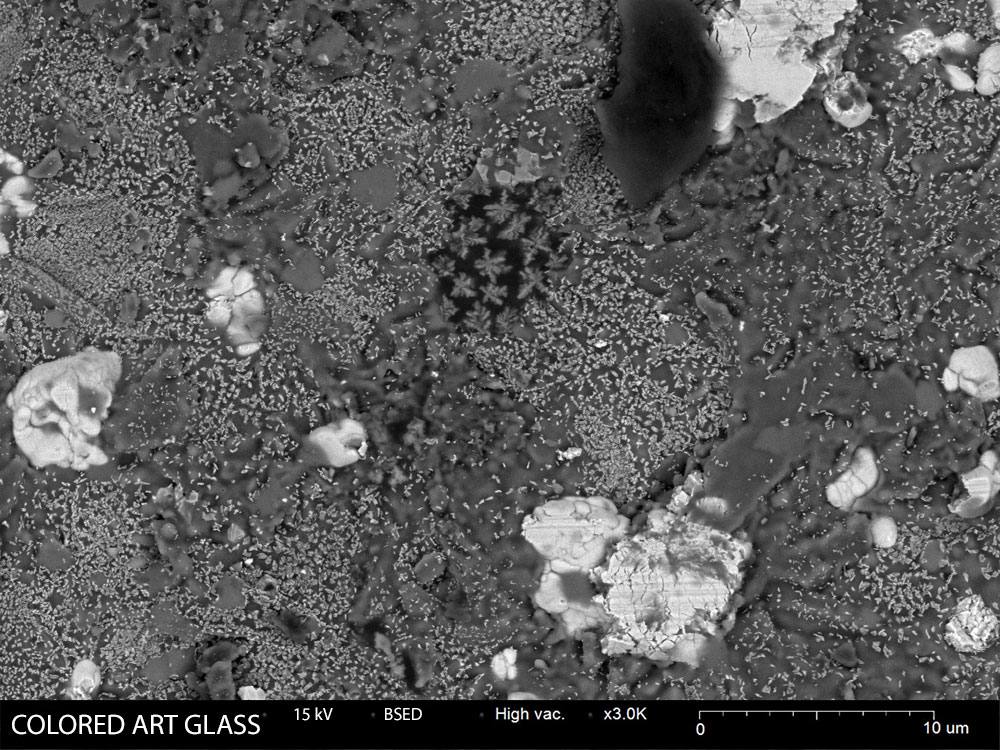
Forensic SEM/EDS begins with scanning the surface of a stub (often a tape lift from a suspect’s hand or clothing) to locate suspect particles. Spherical, micron-scale particles are common GSR indicators, particularly when they contain Pb, Ba, and Sb. This method is robust, widely standardized, and largely automated in forensic laboratories.
Raman Spectroscopy: Unlocking the Organic Side
While SEM handles metals, Raman targets molecular fingerprints. Organic gunpowder components such as nitroglycerin, nitrocellulose, and various nitrates yield distinct Raman spectra. Even post-combustion products or additives specific to certain ammunition brands can be identified. This allows for confirmation of shooting events, particularly in cases involving non-traditional or lead-free ammunition, where metallic markers are reduced or absent.
Improving Accuracy and Confidence
The synergy between SEM and Raman reduces both false positives and false negatives. Particles from brake dust or fireworks might mimic GSR in SEM/EDS, but Raman can confirm the absence of key gunpowder organics, preventing misidentification. Conversely, if a sample contains few metal-rich particles but abundant organic signatures, Raman ensures potential GSR isn’t overlooked.
Non-Destructive, Dual Confirmation
Another critical benefit is preservation of the evidence. Unlike colorimetric chemical tests, Raman spectroscopy is non-destructive and can be performed directly on the same SEM stub. This dual-pathway analysis—elemental via SEM and molecular via Raman—is powerful for courtroom scenarios. It allows analysts to present visual, elemental, and chemical data from the same sample area, enhancing evidentiary weight.
Integrated SEM-Raman Workflow
From Detection to Identification in One Place
In a unified SEM-Raman system, the workflow is streamlined:
- SEM imaging and EDS locate suspect GSR particles.
- Without moving the sample, Raman spectroscopy is performed on those exact particles.
- Correlated software overlays spectral data onto SEM images, tagging each particle with both elemental and molecular information.
This approach reduces the risk of contamination or loss and accelerates time-to-results. It also supports consistent chain-of-custody protocols by maintaining all analysis within a single system and session.
Enhanced Reporting and Clarity
The final forensic report can include a particle’s SEM image, its EDS elemental spectrum, and its Raman chemical fingerprint. This layered evidence paints a compelling picture: not only is the particle morphologically and elementally consistent with GSR, but it also contains molecular traces of gunpowder.
NanoImages’ All-in-One GSR Detection System
NanoImages offers a desktop SEM tailored for forensic applications, equipped with integrated Bruker EDS and Waviks Raman spectroscopy. This compact system allows full GSR analysis from one instrument:
- SEM identifies suspect particles and confirms metal content.
- Raman spectroscopy characterizes associated organics.
- Coordinated optics ensure accurate, co-localized targeting.
- Guided workflows help streamline tasks for users of varying experience levels.
By bringing this level of capability in-house, forensic labs can eliminate the need to shuttle samples between instruments or outsource Raman analysis. Instead, the full GSR profile can be determined rapidly, reliably, and on-site.
Advancing Forensic GSR Analysis
Combining SEM and Raman spectroscopy delivers a forensic toolkit capable of comprehensive GSR detection. It integrates the power of elemental analysis with molecular identification, covering both inorganic primer particles and organic propellant residues. This dual approach improves detection in complex scenarios, supports accurate exclusion of false positives, and enhances evidentiary credibility.
For forensic labs, especially those dealing with modern ammunition types and time-sensitive investigations, integrated SEM-Raman systems represent a significant advancement. By providing robust, reproducible, and multi-dimensional results from a single sample, this technology elevates both the science and the standard of gunshot residue analysis.