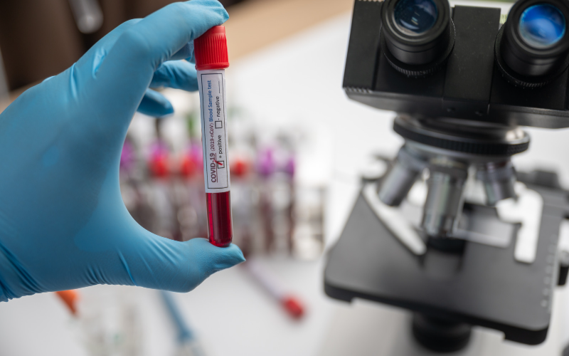
Cells Under Microscope: Exploring Cellular Structures Through Microscopy
Ever wondered how scientists uncover the hidden secrets of cells or how industrial applications benefit from precise cellular analysis? Then you've come to the right place.
Cellular structures hold immense significance across various fields of study, including biology, materials science, and manufacturing. They are the building blocks of life and play a crucial role in understanding diseases, developing new materials, and ensuring the quality of industrial components. To unlock the mysteries of these intricate structures, microscopy has become an indispensable tool.
In this blog, we'll delve into the basics of microscopy, highlighting microscopes commonly used in cellular research. We'll explore the wonders of scanning electron microscopes (SEMs) and how they provide high-resolution imaging of cellular structures.
But why should you care about microscopy? By peering into the microscopic world, we gain valuable insights that help scientists push the boundaries of knowledge and industries optimize their processes. From understanding the intricate machinery within a cell to inspecting the quality of critical components, microscopy plays a vital role in enhancing our understanding of the cellular universe.
So, grab a cup of coffee, sit back, and get ready to discover the hidden wonders beneath the microscope. Our journey is about to begin, where science meets technology, and curiosity leads the way.

The Basics of Microscopy
To truly appreciate the wonders of cellular exploration, we must first grasp the fundamentals of microscopy. Microscopy, in its essence, is the science of magnifying and examining objects that are too small to be seen with the naked eye. By harnessing the power of optics and advanced imaging techniques, microscopes enable us to peer into the intricate details of the microscopic world.
There are several types of microscopes commonly used in cellular research, each with its own strengths and limitations. Let's take a closer look at two primary categories: light microscopy and electron microscopy.
Light Microscopy
Also known as optical microscopy, light microscopy is one of the oldest and most widely used forms of microscopy. It utilizes visible light and a series of lenses to magnify and illuminate the sample. Light microscopes come in various forms, including bright-field, phase-contrast, fluorescence, and confocal microscopy. Each variant offers unique capabilities for visualizing different aspects of cellular structures.
Electron Microscopy
Meanwhile, electron microscopy takes the exploration of cellular structures to a whole new level. Instead of using light, electron microscopes employ a beam of electrons to image the sample with incredibly high resolution. This allows scientists to visualize cellular structures at the nanoscale, revealing intricate details that were previously inaccessible.
Electron microscopy encompasses two main types: transmission electron microscopy (TEM) and scanning electron microscopy (SEM). TEM enables us to observe ultra-thin slices of samples in great detail, while SEM provides a three-dimensional view of the sample surface.
It's important to note that while electron microscopy offers excellent resolution, it requires specialized equipment and sample preparation techniques. Light microscopy, on the other hand, is more accessible and versatile for routine laboratory use.
Now that we have a solid understanding of the different types of microscopes used in cellular research, let's delve deeper into the specifics of SEMs and explore their role in unraveling the mysteries of cellular structures.
SEMs for Cellular Exploration
Among the various microscopy techniques available, SEMs stand out as powerful tools for investigating cellular structures with exceptional detail and clarity. Let’s explore further how they revolutionize our understanding of the microscopic realm.
SEM Overview
SEMs utilize a beam of focused electrons to visualize the surface of a sample in high resolution. Unlike light microscopes, which rely on visible light, SEMs operate on the principles of electron optics. By scanning the sample surface with a finely focused electron beam, SEMs generate detailed images that provide insights into cellular structures at the micro- and nanoscale.
Working Principle
SEMs employ a three-step process to produce images. First, a primary electron beam is emitted from an electron source and focused onto the sample using electromagnetic lenses. The beam then interacts with the sample, causing various signals to be generated. These signals include secondary electrons, backscattered electrons, and X-rays.
Next, detectors capture and measure these signals, translating them into an image that reflects the sample's surface morphology and composition. Finally, the image is displayed on a screen, revealing intricate details of the cellular structures under investigation.
Key Features and Benefits
SEMs are equipped with advanced features and functionalities that make them indispensable tools for cellular exploration. Some notable features include:
- High-resolution Imaging: SEMs deliver exceptional image quality, allowing researchers to observe cellular structures with remarkable clarity.
- User-friendly interface: SEMs are specifically designed with intuitive controls and software, making them accessible to both novice and experienced users.
- Versatility: SEMs accommodate a wide range of samples and provide various imaging modes, enabling researchers to explore different aspects of cellular structures.
- Enhanced productivity: Last but not least, SEMs optimize productivity in cellular research with rapid imaging capabilities and efficient workflows.
Now that we understand the foundations of SEMs and their capabilities, let's dive deeper into the exciting applications of scanning electron microscopes in exploring cellular structures.
SEMs for Cellular Exploration
SEMs have transformed the way we study cellular structures by providing remarkable insights and visualizations at an unprecedented level of detail. Let's explore some of the exciting applications of SEMs in unraveling the mysteries of cellular structures:
Visualizing Surface Morphology
SEMs excel at revealing the surface morphology of cellular structures. By scanning the sample surface with the focused electron beam, SEMs produce highly detailed, three-dimensional images. This enables researchers to observe the intricate surface features of cells, such as microvilli, cilia, or surface texture, with exceptional clarity. Understanding these surface characteristics is crucial in cell biology, tissue engineering, and biomaterials.
Investigating Cellular Interactions and Adhesion
SEMs provide valuable insights into cellular interactions and adhesion processes. By imaging cellular structures at high magnification, researchers can observe the fine details of cellular adhesion complexes, cell-substrate, or cell-cell interactions. This knowledge contributes to a deeper understanding of cellular behavior, cell migration, and tissue development, which have implications in fields ranging from regenerative medicine to cancer research.
Examining Cellular Ultrastructure
SEMs enable the examination of cellular ultrastructure, revealing the internal organization and fine details of cellular components. With sample preparation techniques such as freeze-fracture, critical point drying, or metal coating, SEMs can visualize cellular organelles, cellular membranes, and intracellular structures. This level of detail provides crucial information for studying cellular processes, organelle function, and cellular abnormalities.
Characterizing Biomaterials and Implants
SEMs are vital in characterizing biomaterials and implants used in various biomedical applications. By examining the surface morphology and structure of these materials, SEMs help assess their compatibility with cellular structures, evaluate their biocompatibility, and understand the interactions between materials and cells. This information is invaluable in the development of improved biomaterials, tissue engineering scaffolds, and medical implants.
Advancements in Cellular Analysis Techniques
SEMs continue to evolve with technological advancements, leading to exciting new capabilities in cellular analysis. For example, environmental SEMs (E-SEMs) allow imaging of live cells under controlled environmental conditions, providing insights into dynamic cellular processes. Additionally, SEMs combined with elemental analysis techniques, such as energy-dispersive X-ray spectroscopy (EDS), enable the identification and mapping of elemental composition within cellular structures.
The applications of scanning electron microscopes in exploring cellular structures are vast and impactful. However, SEMs are not the only tools at our disposal. Industrial X-ray inspection systems also contribute significantly to cellular analysis, especially in examining semiconductor components and castings.
Let's explore the realm of X-ray inspection systems and their role in understanding cellular structures further.
X-Ray Inspection Systems for Cellular Analysis
In addition to SEMs, X-ray inspection systems have emerged as valuable tools for cellular analysis. These systems employ X-rays to penetrate samples, providing non-destructive 2D and 3D imaging capabilities.
Let's delve into the world of X-ray inspection systems and their applications in understanding cellular structures, with a specific focus on semiconductor components and castings:
Non-Destructive Cellular Analysis
X-ray inspection systems allow researchers to examine cellular structures without damaging the samples. This non-destructive approach is particularly advantageous when studying delicate or irreplaceable specimens.
These systems generate detailed cross-sectional and volumetric information by capturing X-ray images from different angles, enabling researchers to visualize and analyze cellular structures in three dimensions.
Semiconductor Component Analysis
X-ray inspection systems play a crucial role in the inspection and quality control of semiconductor components. Components like integrated circuits and microchips are intricate structures with distinct features. X-ray systems help identify defects within the semiconductor materials, such as voids, cracks, or delamination. This information is essential for ensuring the functionality and reliability of semiconductor devices.
Casting Inspection
X-ray inspection systems are widely employed in the inspection of castings, which are complex metal components created through the casting process. Cellular structures within castings, such as porosity or inclusions, can significantly affect their mechanical properties and performance.
X-ray inspection systems make detecting and analyzing these internal structures possible, ensuring the quality and integrity of castings used in various industries, including automotive, aerospace, and manufacturing.
Imaging Techniques
X-ray inspection systems utilize different imaging techniques to capture cellular structures within samples. Two commonly used techniques are 2D X-ray radiography and 3D computed tomography (CT). 2D radiography provides detailed 2D images that reveal internal structures, while 3D CT reconstructs a three-dimensional model of the sample, allowing for in-depth analysis of cellular structures in volumetric detail.
SEMs and X-ray inspection systems may have already revolutionized cellular microscopy, but the future promises even more exciting advancements. Let's take a glimpse into the future of cellular microscopy and the possibilities that lie ahead.
Current and Future Perspectives in Cellular Microscopy
As technology continues to evolve at a rapid pace, the future of cellular microscopy holds immense promise. Here are some future perspectives that shape the direction of cellular microscopy:
Advances in Imaging Techniques
Emerging imaging techniques, such as correlative and super-resolution microscopy, will push the boundaries of cellular exploration even further. These techniques will offer unprecedented levels of detail and precision, enabling researchers to unravel cellular structures at the molecular and atomic scales.
Integration of Artificial Intelligence (AI)
Integrating AI algorithms and machine learning into microscopy systems will revolutionize image analysis and data interpretation. AI-powered image processing techniques will enhance automation, accelerate research, and facilitate the extraction of meaningful information from vast datasets, enabling researchers to uncover cellular insights more efficiently.
In Vivo Imaging and Dynamic Observation
Advancements in cellular microscopy aim to capture cellular structures in their natural environment through techniques like intravital microscopy and live-cell imaging. These approaches will enable dynamic observation of cellular processes, providing real-time insights into cell behavior, tissue development, and disease progression.
Multimodal Imaging
The fusion of multiple imaging modalities, such as combining SEM and fluorescence microscopy, will allow researchers to obtain comprehensive information about cellular structures. Multimodal imaging techniques will enable the simultaneous visualization of surface morphology, internal systems, and specific cellular markers, providing a holistic view of cellular complexity.
With these exciting advancements on the horizon, we’re committed to staying at the forefront of cellular microscopy. By embracing emerging technologies and collaborating with researchers, we aim to drive innovation and continue supporting cutting-edge discoveries in cellular analysis.
Conclusion
In this captivating journey through the world of cellular microscopy, we have witnessed the remarkable capabilities of SEMs and X-ray inspection systems in unraveling the mysteries of cellular structures. These tools have revolutionized our understanding of the microscopic realm, from visualizing surface morphology to examining cellular ultrastructure.
SEMs have allowed us to delve into the intricate details of cellular structures, providing high-resolution imaging and valuable insights into cellular interactions, adhesion processes, and biomaterial characterization. On the other hand, X-ray inspection systems have offered non-destructive 2D and 3D analysis, playing a crucial role in inspecting semiconductor components and castings for cellular defects.
Looking ahead, the future of cellular microscopy holds great promise. Technological advancements are set to propel our exploration of cellular structures to new heights. These developments will enable us to unravel the intricacies of cells at the molecular and atomic scales, observe dynamic cellular processes in real time, and obtain comprehensive information through the fusion of different imaging modalities.
As we conclude this captivating exploration, we invite you to join us on the exciting journey of scientific discovery, where technology meets curiosity and every microscopic detail reveals a world of possibilities. Together, let's continue to push the boundaries of cellular microscopy and uncover the intricate beauty within the cells under the microscope.
