Raman Spectroscopy
Correlative Light and Electron Microscopy (CLEM) for molecular fingerprinting.
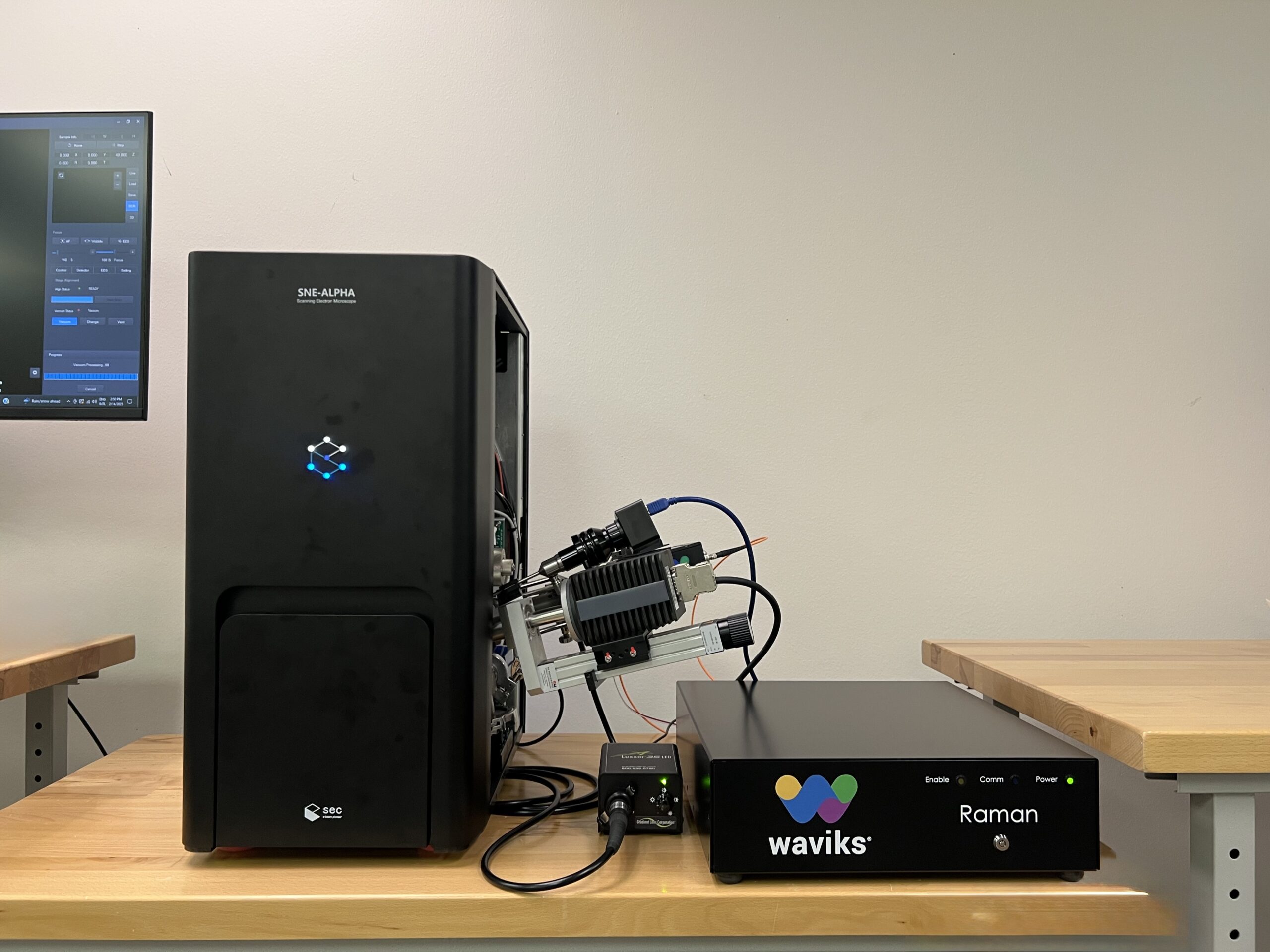
Our CLEM-enabled Raman system reveals molecular structure and chemical bonding information that complements SEM imaging and EDS elemental analysis. Perform simultaneous correlative light and electron microscopy to identify polymers, minerals, pharmaceuticals, and organic compounds with high specificity.
See Raman-SEM in Action
Why CLEM-Enabled Raman + SEM?
True CLEM Integration
Correlative Light and Electron Microscopy in one instrument. Acquire SEM images and Raman spectra from the exact same location simultaneously.
Molecular Identification
Identify polymers, minerals, and organic compounds by their unique vibrational fingerprints. Distinguish materials that look identical in SEM.
CLEM Chemical Mapping
Create spatial distribution maps of molecular species with CLEM precision. Correlate optical and electron data for complete characterization.
Stress Analysis
Detect residual stress in crystalline materials through Raman peak shifts. Non-destructive CLEM stress mapping.
Waviks Vesta™ Raman System Specifications
Simultaneous Correlated Light and Electron Microscopy (CLEM) in the SEM
Optical Performance
System Integration
Key Features
- Simultaneous Correlated Light and Electron (CLEM) microscopy
- Combine SEM imaging, EDS analysis, and Raman in one workflow
- Non-contact sample heating with no high temperature limit
- Temperature-dependent EBSD when combined with EBSD detector
- High-mag and large field-of-view imaging for quick alignment
- Modular design for easy integration of multiple Vesta™ probes
- Quick-change filter box for rapid wavelength switching
- Python SDK and Ethernet link for automation
Applications
Materials Science
Metals, polymers, composites, failure analysis.
Life Sciences
Cells, tissues, biological structures.
Semiconductors
Wafer inspection, defect analysis, IC packaging.
Forensics
GSR analysis, trace evidence, fiber comparison.
Geosciences
Minerals, rocks, paleontology.
Nanomaterials
Nanoparticles, thin films, quantum dots.
Related Products
Resources
Experience True CLEM Capability
Correlative Light and Electron Microscopy: SEM imaging, EDS analysis, and Raman spectroscopy in one integrated workflow.
Discuss CLEM Integration