
GeoSciences
Mineral identification, rock analysis, petrography, and geological sample characterization
Scanning electron microscopy is indispensable for geological research, providing high-resolution imaging and elemental analysis of minerals, rocks, and sediments. SEM-EDS enables rapid mineral identification, textural analysis, and compositional mapping that complements traditional optical petrography.
The SNE-Alpha desktop SEM brings geological analysis to field stations, teaching labs, and core facilities. Its ability to image unpolished samples and analyze at low vacuum makes it particularly suited for rapid mineral identification and screening applications.
Key Geoscience Applications
- Mineral Identification: Combine morphology with EDS chemistry for rapid mineral ID. Distinguish polymorphs and identify trace phases
- Petrography: Examine rock textures, crystal habits, and mineral relationships at microscale resolution
- Sedimentology: Characterize grain morphology, surface textures, and diagenetic features in sediments and sedimentary rocks
- Ore Microscopy: Identify ore minerals, analyze textures, and assess liberation characteristics
- Micropaleontology: Image microfossils including foraminifera, ostracods, and conodonts
- Clay Mineralogy: Characterize clay mineral morphology and composition in soils and sediments
Sample Requirements
Geological samples are well-suited for desktop SEM analysis:
- Rock Chips: Fresh fracture surfaces reveal crystal morphology and mineral relationships
- Polished Sections: Standard thin section or polished mounts for quantitative EDS analysis
- Loose Grains: Sediment samples mounted on carbon tape for morphological studies
- Core Samples: Small pieces from drill cores for rapid screening
Most geological samples are naturally conductive or semi-conductive, requiring minimal preparation. Non-conductive samples can be carbon coated or imaged in variable pressure mode.
Geoscience SEM Images
Sample images captured with the SNE-Alpha desktop SEM.
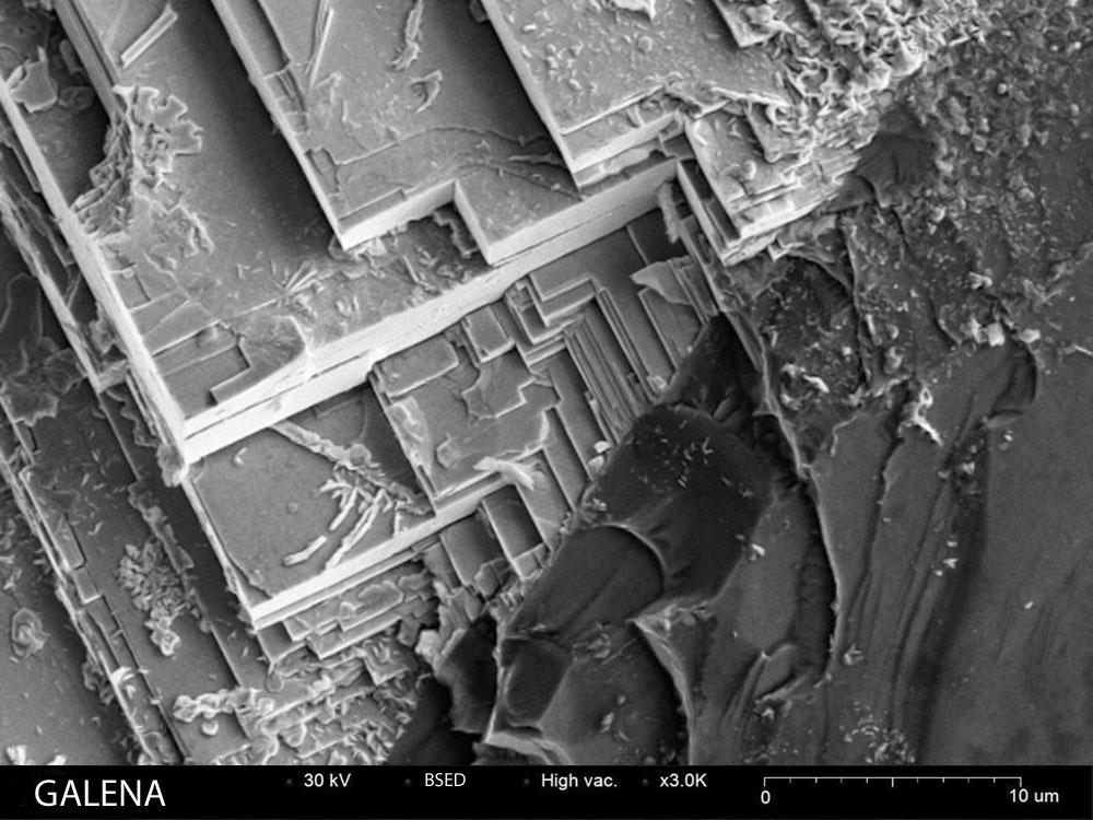
Galena
Lead sulfide mineral showing characteristic cubic crystal structure
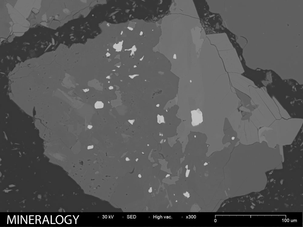
Mineral Sample
Complex mineral assemblage showing phase relationships
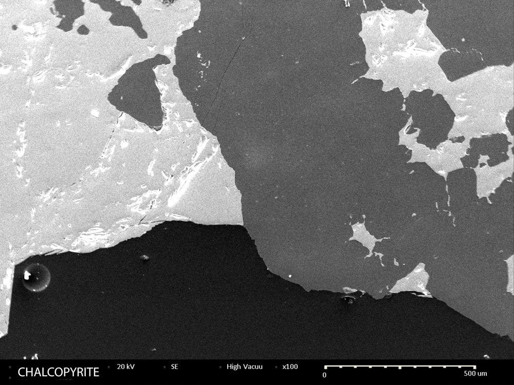
Chalcopyrite
Copper iron sulfide ore mineral with surface texture detail
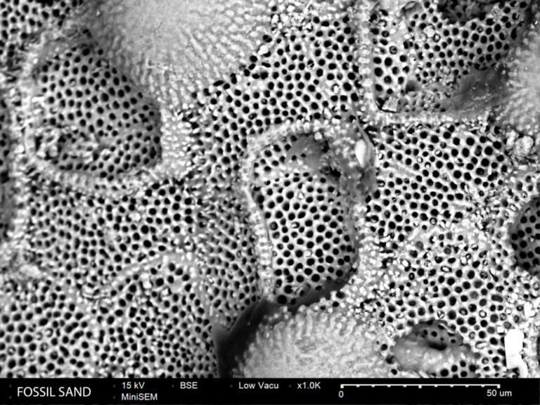
Fossil Sand
Fossiliferous sand grains showing microfossil content
Analytical Techniques
SE Imaging
Topographic imaging reveals crystal morphology, surface textures, and microstructural features.
BSE Imaging
Atomic number contrast distinguishes mineral phases in polished sections for modal analysis.
EDS Mapping
Element distribution maps show zonation patterns and phase relationships in complex assemblages.
Point Analysis
Quantitative mineral chemistry for classification and provenance studies.
Recommended Equipment
SNE-Alpha Desktop SEM
Compact, high-resolution scanning electron microscope ideal for research and quality control applications.
Bruker XFlash EDS
Energy dispersive X-ray spectroscopy for elemental analysis and material identification.
MCM-100 Sputter Coater
Prepare non-conductive samples with gold or platinum coatings for optimal SEM imaging.
Related Applications
Resources
Geological Analysis Solutions
See how the SNE-Alpha can support your geological research and teaching programs.
Request Sample Analysis