
Forensic Science
GSR analysis, fiber comparison, trace evidence, and crime scene investigation
The SNE-Alpha desktop SEM is used by forensic labs for gunshot residue (GSR) analysis, trace evidence examination, fracture matching, and document forgery investigation. Its 90-second vacuum and compact footprint make it deployable in crime labs without specialized infrastructure. Combine with EDS for elemental confirmation of GSR particles. See full SNE-Alpha specifications →
Scanning electron microscopy combined with energy dispersive spectroscopy (SEM-EDS) is an essential tool in forensic laboratories worldwide. The technique provides both morphological imaging and elemental analysis of trace evidence, allowing examiners to identify and compare materials with high specificity.
The SNE-Alpha desktop SEM with integrated Bruker EDS brings forensic-grade analysis to police departments, crime labs, and educational institutions. Its compact size and ease of operation make it accessible for routine casework while delivering the resolution and analytical capabilities demanded by forensic standards.
Key Forensic Applications
- Gunshot Residue (GSR) Analysis: Detect and identify characteristic particles containing lead, barium, and antimony. Automated particle search locates GSR particles efficiently
- Fiber Analysis: Compare fiber morphology, surface characteristics, and cross-sectional shapes. Distinguish natural from synthetic fibers
- Paint Chip Comparison: Examine layer structure, pigment particles, and surface texture. Match chips to known sources
- Tool Mark Evidence: Image striations and impressions on metals, plastics, and other materials at high magnification
- Glass Analysis: Characterize glass fragment morphology and use EDS for elemental profiling
- Document Examination: Analyze inks, papers, and printing methods. Detect alterations and authenticate documents
GSR Analysis with SEM-EDS
Gunshot residue analysis remains one of the most important forensic applications of SEM-EDS. When a firearm is discharged, the primer produces microscopic particles that deposit on nearby surfaces and skin. These particles have characteristic spherical morphology and contain specific elemental combinations:
- Lead (Pb): From the primer and bullet
- Barium (Ba): From barium nitrate in the primer
- Antimony (Sb): From antimony sulfide in the primer
Particles containing all three elements are considered characteristic of GSR and provide strong evidence of firearm discharge.
Forensic SEM Images
Sample images captured with the SNE-Alpha desktop SEM.
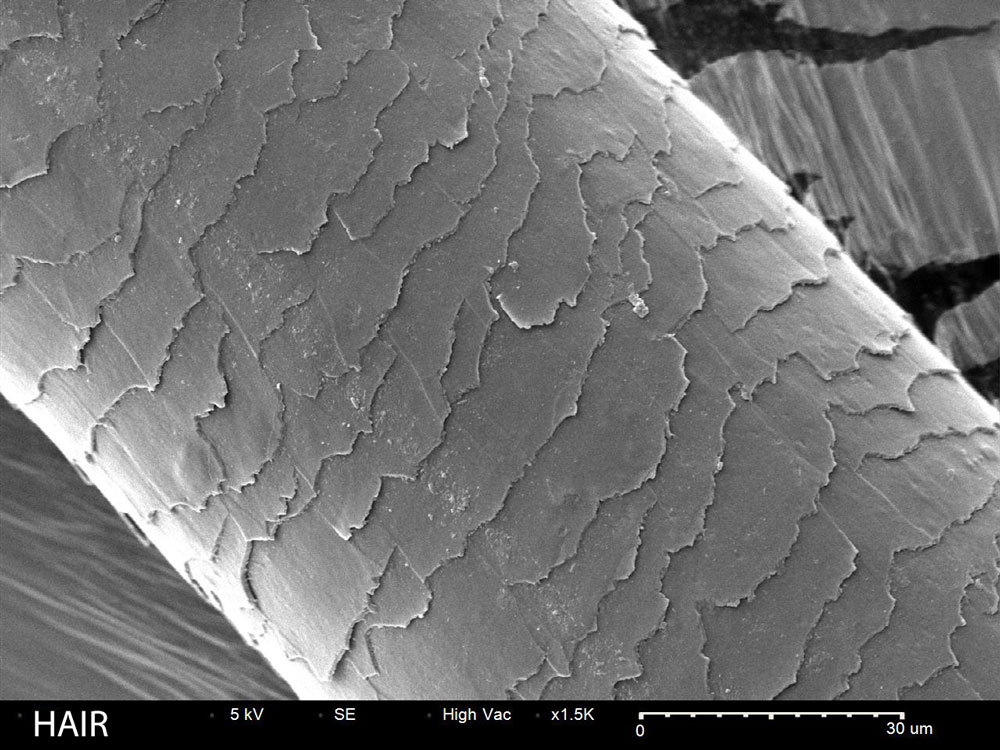
Human Hair
Hair shaft showing cuticle scale pattern for comparison analysis
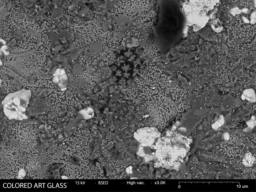
Glass Fragments
Colored glass particles for forensic trace evidence analysis
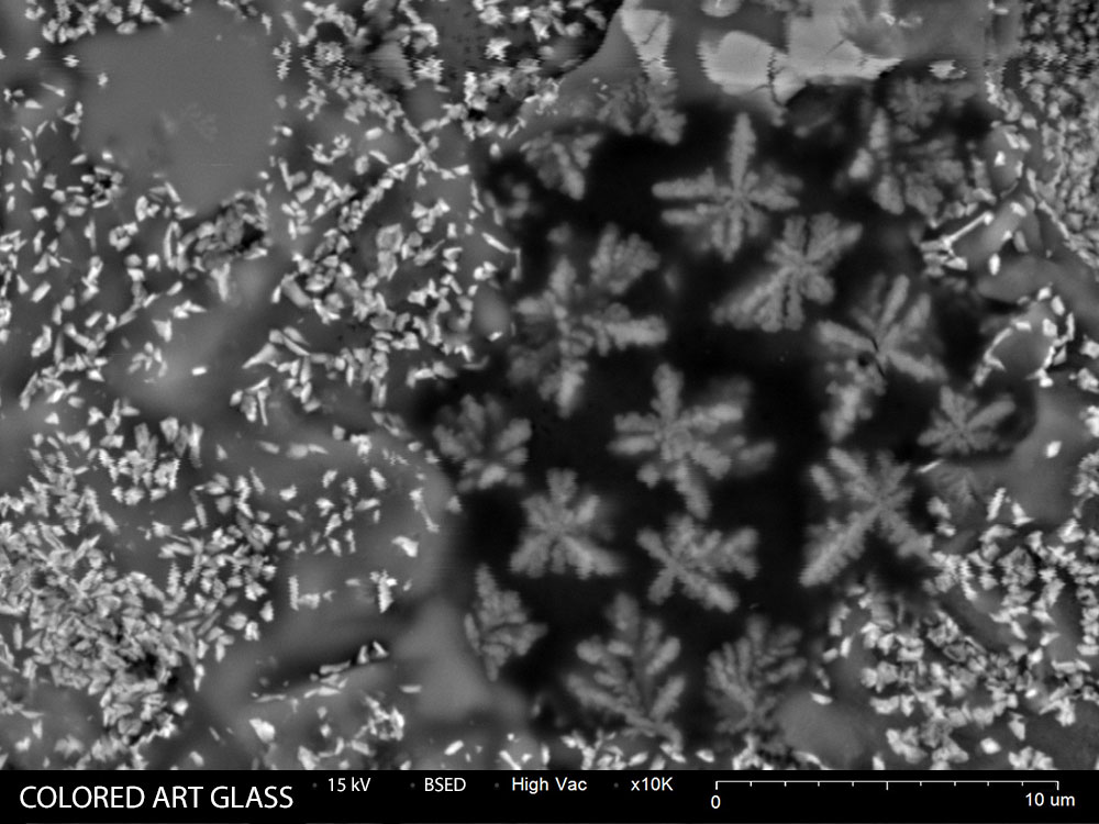
Glass Fragment Detail
High magnification view of glass fracture surface
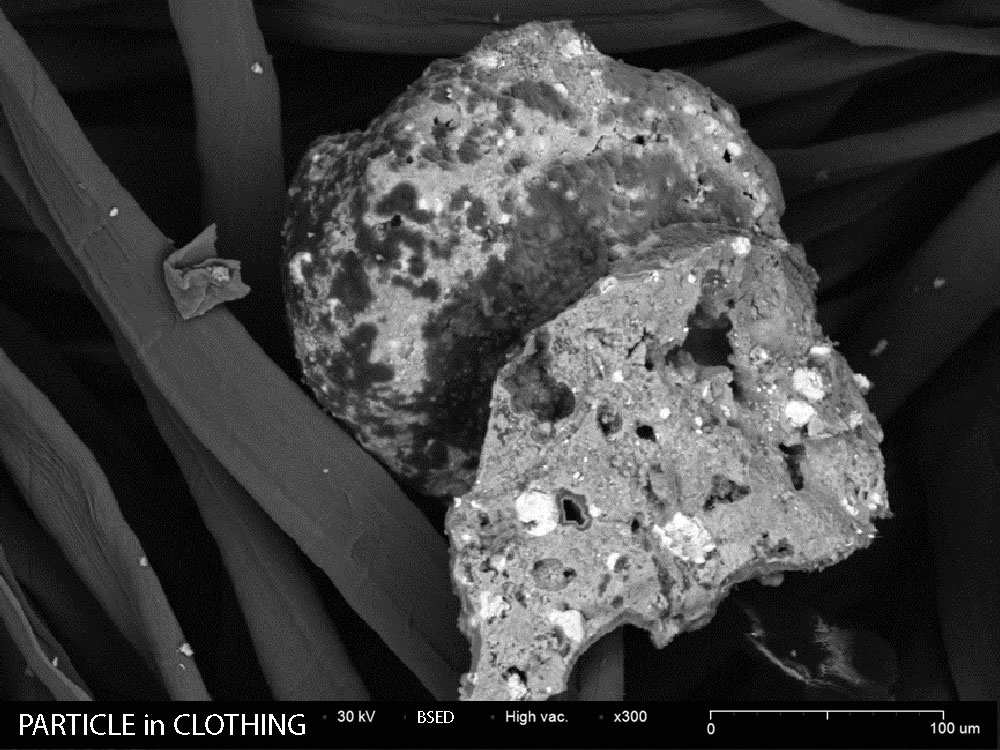
Particle in Clothing
Trace evidence particle found embedded in textile fibers
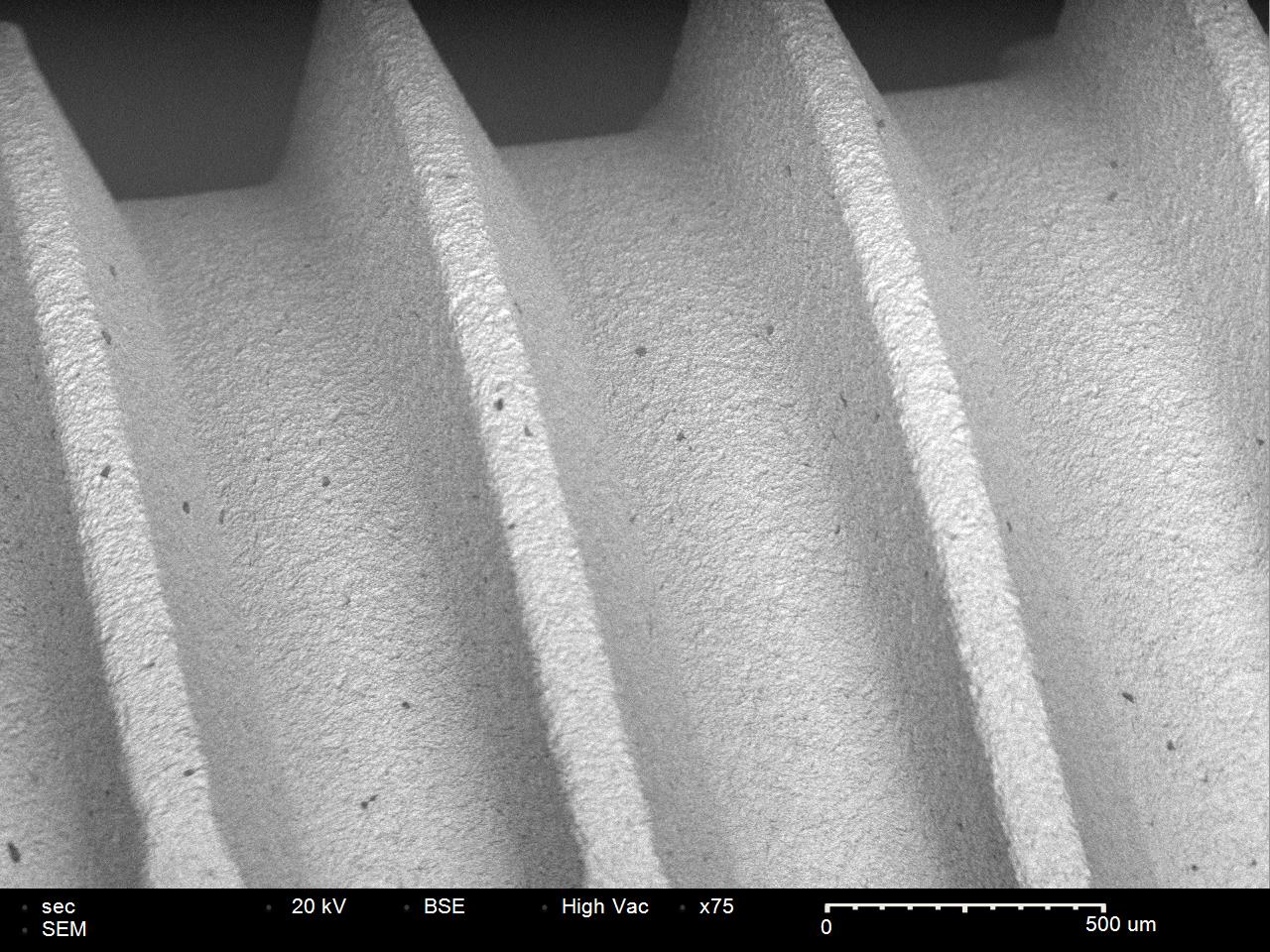
Metal Fastener
Surface detail and tool marks on metal fastener
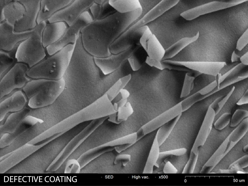
Defective Coating
Surface coating defect analysis for failure investigation
Analytical Techniques
SE Imaging
High-resolution topographic imaging reveals surface features, textures, and morphology for visual comparison.
BSE Imaging
Backscatter electron imaging provides compositional contrast to distinguish materials with different atomic numbers.
EDS Analysis
Elemental identification and quantification for GSR, glass, paint, and other trace evidence.
Automated Particle Search
Software-automated scanning locates particles of interest (like GSR) across large sample areas.
Recommended Equipment
SNE-Alpha Desktop SEM
Compact, high-resolution scanning electron microscope ideal for research and quality control applications.
Bruker XFlash EDS
Energy dispersive X-ray spectroscopy for elemental analysis and material identification.
MCM-100 Sputter Coater
Prepare non-conductive samples with gold or platinum coatings for optimal SEM imaging.
Related Applications
Resources
Forensic Analysis Solutions
See how the SNE-Alpha can support your crime lab or forensic science program.